Electron microscopy techniques

Scanning electron microscope

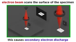
A scanning electron microscope (SEM) is a type of electron microscope that produces images of a sample by scanning the surface with a focused beam of electrons. The electrons interact with atoms in the sample, producing various signals that contain information about the surface topography and composition of the sample. The electron beam is scanned in a raster scan pattern, and the position of the beam is combined with the intensity of the detected signal to produce an image. In the most common SEM mode, secondary electrons emitted by atoms excited by the electron beam are detected using a secondary electron detector (Everhart–Thornley detector). The number of secondary electrons that can be detected, and thus the signal intensity, depends, among other things, on specimen topography. Some SEMs can achieve resolutions better than 1 nanometer. Specimens are observed in high vacuum in a conventional SEM, or in low vacuum or wet conditions in a variable pressure or environmental SEM, and at a wide range of cryogenic or elevated temperatures with specialized instruments. (Wikipedia).

Scanning electron microscope
Video thumbnail

DIY Scanning Electron Microscope - Overview

Today, I finally produced an image with my DIY scanning electron microscope. I've spent the last few months working on this project, and am encouraged by today's success. There is still a lot of work left to do in making the image higher resolution, and eliminating sources of noise, howe

From playlist Scanning Electron Microscope

Video thumbnail

DIY Scanning Electron Microscope - Electron Gun Detail

I explain the detailed operation of the electron gun in my DIY scanning electron microscope project.

From playlist Scanning Electron Microscope

Video thumbnail

DIY Scanning Electron Microscope - Operation procedure

After getting back from Maker Faire (which is always a hugely enjoyable and inspiring event), I thought that my microscope might need some repairs. As it turned out, I only had to change the filament and tighten some screws that came loose during the trip back from the Faire. The microsc

From playlist Scanning Electron Microscope

Video thumbnail

Electron beam control in a scanning electron microscope

I describe electron beam optics and deflection as they are employed in scanning electron microscopy. http://web.jfet.org/vclk/ - Deflection amplifier http://www.johngineer.com/blog/?p=648 - CRT Christmas tree http://www.fei.com/ - FEI Company

From playlist Scanning Electron Microscope

Video thumbnail

DIY Scanning Electron Microscope - Sources, Costs and References

Please visit my blog post to see the references and sources for this project: http://benkrasnow.blogspot.com/2011/03/diy-scanning-electron-microscope_26.html

From playlist Scanning Electron Microscope

Video thumbnail

The Electron Microscope: Where did it come from? | Stuff of Genius

Once we exhausted the ability to magnify objects through the use of light and lenses, we had to look for a new method. Enter the electron microscope. A beam of electrons can get in more of the nooks and crannies of matter to see more detail. Stuff of Genius tells the story behind everyday

From playlist Gizmos & Widgets

Video thumbnail

Electron Microscopy (TEM and SEM)

We've talked a lot about light microscopy, but this technique has inherent limitations in resolution and magnification. The next paradigm in microscopy that emerged in the middle of the 20th century was electron microscopy. Because electrons have much shorter wavelengths than photons, elec

From playlist Microbiology/Infectious Diseases

Video thumbnail

DIY Scanning Electron Microscope - Tektronix contest entry

Vote for me here: http://mytektronixscope.com/videos/ I am using my Tektronix 2246 analog oscilloscope to show the image generated by my DIY electron microscope. The 2246 is operating in X-Y mode, with the channels connected to a raster scan generator. The vertical scanning frequency

From playlist Scanning Electron Microscope

Video thumbnail

Peng Wang - Electron Ptychography: Emerging Computational Microscopy for Physical/Biological Science

Recorded 28 October 2022. Peng Wang of the University of Warwick presents "Electron Ptychography: An Emerging Computational Microscopy for Physical and Biological Sciences" at IPAM's Mathematical Advances for Multi-Dimensional Microscopy Workshop. Abstract: Ptychography is an emerging comp

From playlist 2022 Mathematical Advances for Multi-Dimensional Microscopy

Video thumbnail

Electron microscope image capture with an oscilloscope

T-shirt: http://www.teespring.com/AppliedScience 11:10 - Skip directly to seeing the collected images I show how I collect digital electron microscope images with my Tek MDO3000 oscilloscope.

From playlist Electronics

Video thumbnail

Viewing an active electronic circuit with a scanning electron microscope

I used my DIY scanning electron microscope to view a 555 timer circuit while it was powered. The circuit is a simple oscillator with a very long time constant to make the changes easy to see. My plan was to view the silicon die itself, and hopefully discern changes in its internal circuitr

From playlist Electronics

Video thumbnail

Nigel Browning - Inpainting Approaches to Dose Control in High Resolution and In-Situ STEM

Recorded 24 October 2022. Nigel Browning of the University of Liverpool presents "Inpainting Approaches to Dose Control in High Resolution and In-Situ STEM" at IPAM's Mathematical Advances for Multi-Dimensional Microscopy Workshop. Abstract: For many imaging and microanalysis experiments u

From playlist 2022 Mathematical Advances for Multi-Dimensional Microscopy

Video thumbnail

Mod-01 Lec-01 Introduction to Nanotechnology

Nano structured materials-synthesis, properties, self assembly and applications by Prof. A.K. Ganguli,Department of Nanotechnology,IIT Delhi.For more details on NPTEL visit http://nptel.ac.in

From playlist IIT Delhi: Nano structured materials-synthesis, properties, self assembly and applications | CosmoLearning.org Materials Science

Video thumbnail

Materialism Podcast Ep 10. Seeing with Electrons

The optical microscope was discovered in the 17th century and ever since has been a staple of scientific discovery. It uncovered germs and let humanity investigate the microscopic world. However, optical microscopes face a limit to their resolution since they rely on visible light. If mate

From playlist Materialism Podcast

Related pages

Charge carrier | Liquid-Phase Electron Microscopy | Focused ion beam | Triangulation (computer vision) | Electron shell | Electronvolt | Transmission electron microscopy | Cathodoluminescence | Ion | Immunogold labelling | Order of magnitude | Electron gun | Electron | Nanometre | Torr | Optimal estimation | Pixel | Secondary electrons | Phase transition | Scanning transmission electron microscopy | Digital image | Energy | Electron hole | Electron microscope | Annular dark-field imaging | Atomic number | Osmium tetroxide | Ion beam