https://www.patreon.com/edmundsj If you want to see more of these videos, or would like to say thanks for this one, the best way you can do that is by becoming a patron - see the link above :). And a huge thank you to all my existing patrons - you make these videos possible. In this video
From playlist Optoelectronic and Photonic Devices

Particle Physics (35 of 41) What is a Photon? 19. The Magnetic Field Effect
Visit http://ilectureonline.com for more math and science lectures! In this video I will explain the effects of the magnetic field in electromagnetic radiation. Next video in the Particle Physics series can be seen at: http://youtu.be/RurZpNnnwds
From playlist PHYSICS 65 PARTICLE PHYSICS

Physics - Ch 66 Quantum Mechanics 2: Basic Concepts (2 of 38) How are Photons Absorbed? 1
Visit http://ilectureonline.com for more math and science lectures! In this video I will explain how photons are absorbed by matter. Next video in this series can be seen at: https://youtu.be/tk2qqRuSlCg
From playlist PHYSICS 66 - QUANTUM MECHANICS


Sergei Kalinin: "Deep Learning Dive into the Scanning Transmission Electron Microscopy: Material..."
Machine Learning for Physics and the Physics of Learning 2019 Workshop II: Interpretable Learning in Physical Sciences "Deep Learning Dive into the Scanning Transmission Electron Microscopy: Materials Design, Learning Physics, and Atomic Manipulation" Sergei Kalinin, Oak Ridge National La
From playlist Machine Learning for Physics and the Physics of Learning 2019

Petar Petrov - Laser phase-contrast transmission electron microscopy & computational opportunities
Recorded 15 November 2022. Petar Petrov of the University of California, Berkeley, presents "Laser phase-contrast transmission electron microscopy and associated computational opportunities" at IPAM's Cryo-Electron Microscopy and Beyond Workshop. Abstract: A phase plate can provide optimum
From playlist 2022 Cryo-Electron Microscopy and Beyond

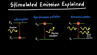
The physics of a laser - how it works. How the atom interacts with light. I’ll use this knowledge to simulate a working laser. We will learn how LASERs relies on Stimulated absorption, Spontaneous emission, and most importantly: Stimulated Emission- This last type interacts with an excite
From playlist New to the channel? Try these
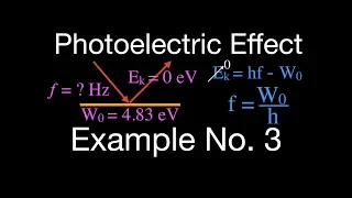
Photoelectric Effect (4 of 8) Example No.3
Includes one easy to follow worked example for the photoelectric effect showing how to determine the minimum frequency and wavelength of light needed to emit an electron from a metal with a know work function. You can see a listing of all my videos at my website, http://www.stepbystepscien
From playlist Quantum Mechanics

Neural Circuit Dynamics During Virtual Navigation and Decision-Making - David Tank
David Tank, Professor of neuroscience and molecular biology at Princeton University, focused on the mechanisms of persistent neural activity and the development and application of rodent virtual reality systems, large-scale optical recording and electrophysiology to study neural circuit dy
From playlist Wu Tsai Neurosciences Institute
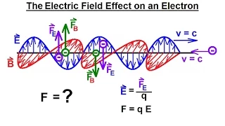
Particle Physics (36 of 41) What is a Photon? 20. The Electric Field
Visit http://ilectureonline.com for more math and science lectures! In this video I will explain the effects of the electric field oscillation on the electron. Next video in the Particle Physics series can be seen at: http://youtu.be/6-C3yE78hZg
From playlist PHYSICS 65 PARTICLE PHYSICS

Chris Jacobsen - Coherent x-ray imaging: how big can we go small? - IPAM at UCLA
Recorded 12 October 2022. Chris Jacobsen of the Argonne National Laboratory/Northwestern University presents "Coherent x-ray imaging: how big can we go small?" at IPAM's Diffractive Imaging with Phase Retrieval Workshop. Abstract: Coherent x-ray imaging methods such as ptychography are dev
From playlist 2022 Diffractive Imaging with Phase Retrieval - - Computational Microscopy
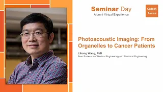
Photoacoustic Imaging: From Organelles to Cancer Patients / Seminar Day, Session III
Photoacoustic Imaging: From Organelles to Cancer Patients / Seminar Day, Session III Saturday, May 15, 2021 12:30 PM Using a combination of light and sound, Lihong Wang is noninvasively peering deeper inside biological tissues than previously possible. Hear about how his novel imaging t
From playlist Seminar Day
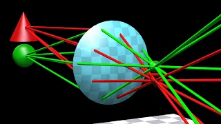
Lenses, refraction, and optical illusions of light
Optics, lenses, and optical illusions created by the refraction of light explained with 3D ray diagrams. My Patreon page is at https://www.patreon.com/EugeneK
From playlist Physics
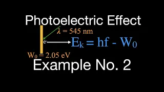
Photoelectric Effect (3 of 8 ) Example No.2 (Very Helpful)
Includes one very helpful worked example for the photoelectric effect for how to calculate the kinetic energy and velocity of the photoelectrons. The photoelectric effect is the emission of electrons when light is shined on a material. Electrons emitted in this way can be called photoelec
From playlist Quantum Mechanics

MIT 7.016 Introductory Biology, Fall 2018 Instructor: Adam Martin View the complete course: https://ocw.mit.edu/7-016F18 YouTube Playlist: https://www.youtube.com/playlist?list=PLUl4u3cNGP63LmSVIVzy584-ZbjbJ-Y63 Professor Martin introduces cell imaging techniques, which are tools that all
From playlist MIT 7.016 Introductory Biology, Fall 2018
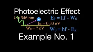
Photoelectric Effect (2 of 8) Example No.1 (Easy to Follow)
Includes one easy to follow worked example for the photo electric effect on how to calculate the work function. The photoelectric effect is the emission of electrons when light is shined on a material. Electrons emitted in this way can be called photoelectrons. In 1905, Albert Einstein p
From playlist Quantum Mechanics
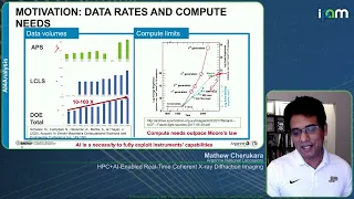
Mathew Cherukara - HPC+AI-Enabled Real-Time Coherent X-ray Diffraction Imaging - IPAM at UCLA
Recorded 14 October 2022. Mathew Cherukara of Argonne National Laboratory presents "HPC+AI-Enabled Real-Time Coherent X-ray Diffraction Imaging" at IPAM's Diffractive Imaging with Phase Retrieval Workshop. Abstract: he capabilities provided by next generation light sources such as the Adva
From playlist 2022 Diffractive Imaging with Phase Retrieval - - Computational Microscopy

The Technology of Optical Superoscillations by Nikolay I Zheludev
DISCUSSION MEETING STRUCTURED LIGHT AND SPIN-ORBIT PHOTONICS ORGANIZERS: Bimalendu Deb (IACS Kolkata, India), Tarak Nath Dey (IIT Guwahati, India), Subhasish Dutta Gupta (UOH, TIFR Hyderabad, India) and Nirmalya Ghosh (IISER Kolkata, India) DATE: 29 November 2022 to 02 December 2022 VE
From playlist Structured Light and Spin-Orbit Photonics - Edited
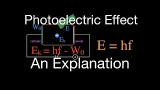
Photoelectric Effect (1 of 8) An Explanation (Clear and Simple)
Explains how the photoelectric effect works. Also includes one worked example for how to calculate the kinetic energy and velocity of the photoelectrons. The photoelectric effect is the emission of electrons when light is shined on a material. Electrons emitted in this way can be called p
From playlist Quantum Mechanics
Paul Weiss - Leveraging Sparsity in Scanning Probe Microscopy - IPAM at UCLA
Recorded 15 September 2022. Paul Weiss of the University of California, Los Angeles, presents "Leveraging Sparsity in Scanning Probe Microscopy" at IPAM's Computational Microscopy Tutorials. Learn more online at: http://www.ipam.ucla.edu/programs/workshops/computational-microscopy-tutorial
From playlist Tutorials: Computational Microscopy 2022