Electron microscopy techniques

High-resolution transmission electron microscopy

High-resolution transmission electron microscopy is an imaging mode of specialized transmission electron microscopes that allows for direct imaging of the atomic structure of samples. It is a powerful tool to study properties of materials on the atomic scale, such as semiconductors, metals, nanoparticles and sp2-bonded carbon (e.g., graphene, C nanotubes). While this term is often also used to refer to high resolution scanning transmission electron microscopy, mostly in high angle annular dark field mode, this article describes mainly the imaging of an object by recording the two-dimensional spatial wave amplitude distribution in the image plane, in analogy to a "classic" light microscope. For disambiguation, the technique is also often referred to as phase contrast transmission electron microscopy. At present, the highest point resolution realised in phase contrast transmission electron microscopy is around 0.5 ångströms (0.050 nm). At these small scales, individual atoms of a crystal and its defects can be resolved. For 3-dimensional crystals, it may be necessary to combine several views, taken from different angles, into a 3D map. This technique is called electron crystallography. One of the difficulties with high resolution transmission electron microscopy is that image formation relies on phase contrast. In phase-contrast imaging, contrast is not intuitively interpretable, as the image is influenced by aberrations of the imaging lenses in the microscope. The largest contributions for uncorrected instruments typically come from defocus and astigmatism. The latter can be estimated from the so-called Thon ring pattern appearing in the Fourier transform modulus of an image of a thin amorphous film. (Wikipedia).

High-resolution transmission electron microscopy
Video thumbnail

Carl Zeiss S-planar lens pt.2: resolution explained

In this video we take a closer look at the maximum imaging resolution of the Carl Zeiss S-Planar lens. Assuming it satisfies the Rayleigh limit, the maximum resolution of this lens should be around 723nm or 0.7um according to theory. Measurements show that the resolution is actually somewh

From playlist Carl Zeiss S-planar Photolithography Lens

Video thumbnail

Electron Microscopy (TEM and SEM)

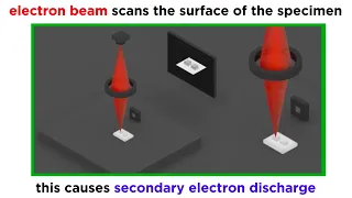
We've talked a lot about light microscopy, but this technique has inherent limitations in resolution and magnification. The next paradigm in microscopy that emerged in the middle of the 20th century was electron microscopy. Because electrons have much shorter wavelengths than photons, elec

From playlist Microbiology/Infectious Diseases

Video thumbnail

Physics - Optics: Circular Aperture - Angle of Resolution (3 of 6) Resolution Power of the Human Eye

Visit http://ilectureonline.com for more math and science lectures! In this video I will explain the resolution ability of the human eye.. Next video in series: http://youtu.be/H2YJYFXo3yo

From playlist PHYSICS 61 DIFFRACTION OF LIGHT

Video thumbnail

Physics - Optics: Circular Aperture - Angle of Resolution (5 of 6) Resolution of the Hubble

Visit http://ilectureonline.com for more math and science lectures! In this video I will find the resolution of the Hubble Space Telescope. Next video in series: http://youtu.be/yFFW20YnGsQ

From playlist PHYSICS 61 DIFFRACTION OF LIGHT

Video thumbnail

Mary Scott - Supervised and Unsupervised approaches for Electron Microscopy Data Analysis

Recorded 01 December 2022. Mary Scott of the University of California, Berkeley, presents "Supervised and Unsupervised approaches for Electron Microscopy Data Analysis" at IPAM's Multi-Modal Imaging with Deep Learning and Modeling Workshop. Abstract: Recently, materials science has undergo

From playlist 2022 Multi-Modal Imaging with Deep Learning and Modeling

Video thumbnail

Peng Wang - Electron Ptychography: Emerging Computational Microscopy for Physical/Biological Science

Recorded 28 October 2022. Peng Wang of the University of Warwick presents "Electron Ptychography: An Emerging Computational Microscopy for Physical and Biological Sciences" at IPAM's Mathematical Advances for Multi-Dimensional Microscopy Workshop. Abstract: Ptychography is an emerging comp

From playlist 2022 Mathematical Advances for Multi-Dimensional Microscopy

Video thumbnail

Spectrum of Hg Lamp / amazing science experiment

Identify the spectral lines of Hg lamp Enjoy the amazing colors! Music: https://www.bensound.com/

From playlist Optics

Video thumbnail

Physics 59 Optical Instruments (10 of 20) Magnification of a Microscope: Exact Approach

Visit http://ilectureonline.com for more math and science lectures! To donate: http://www.ilectureonline.com/donate https://www.patreon.com/user?u=3236071 We will learn the EXACT approach to find the magnification of a microscope given the length of microscope=20cm, focal distance object

From playlist PHYSICS 59 OPTICAL INSTRUMENTS

Video thumbnail

Physics - Optics: Diffraction Grating (7 of 7) Rayleigh's Criteria for Resolution

Visit http://ilectureonline.com for more math and science lectures! In this video I will discuss Rayleigh's criteria for resolution. First video in series: http://youtu.be/-pki5mp0wm4

From playlist PHYSICS 61 DIFFRACTION OF LIGHT

Video thumbnail

Microscopes: optical vs SEM vs TEM vs AFM

In order to examine defects and imperfections in materials, we need microscopes capable of enhancing our vision beyond the human eye capability. To access the micro, nano, and atomic scales needed we must rely on different microscopes. The microscope options vary immensely in terms of cost

From playlist Materials Sciences 101 - Introduction to Materials Science & Engineering 2020

Video thumbnail

Mod-01 Lec-37 Transmission Electron Microscopy

Nanostructures and Nanomaterials: Characterization and Properties by Characterization and Properties by Dr. Kantesh Balani & Dr. Anandh Subramaniam,Department of Nanotechnology,IIT Kanpur.For more details on NPTEL visit http://nptel.ac.in.

From playlist IIT Kanpur: Nanostructures and Nanomaterials | CosmoLearning.org

Video thumbnail

Petar Petrov - Laser phase-contrast transmission electron microscopy & computational opportunities

Recorded 15 November 2022. Petar Petrov of the University of California, Berkeley, presents "Laser phase-contrast transmission electron microscopy and associated computational opportunities" at IPAM's Cryo-Electron Microscopy and Beyond Workshop. Abstract: A phase plate can provide optimum

From playlist 2022 Cryo-Electron Microscopy and Beyond

Video thumbnail

Sarah Haigh - Probing atomic behaviour in liquids with STEM : opportunities for machine learning

Recorded 30 November 2022. Sarah Haigh of the University of Manchester presents "Probing atomic behaviour in liquids with STEM : opportunities for machine learning" at IPAM's Multi-Modal Imaging with Deep Learning and Modeling Workshop. Learn more online at: http://www.ipam.ucla.edu/progra

From playlist 2022 Multi-Modal Imaging with Deep Learning and Modeling

Video thumbnail

Alberto Bartesaghi - Imaging biomolecules & high-resolution single-particle cryo-electron microscopy

Recorded 13 October 2022. Alberto Bartesaghi of Duke University presents "Imaging biomolecules using high-resolution single-particle cryo-electron microscopy" at IPAM's Diffractive Imaging with Phase Retrieval Workshop. Abstract: Single-particle cryo-electron microscopy (EM) is a popular t

From playlist 2022 Diffractive Imaging with Phase Retrieval - - Computational Microscopy

Video thumbnail

Juan Carlos Idrobo - Quantum Phenomena & Electron Microscopy: New Possibilities & Limitations

Recorded 28 October 2022. Juan-Carlos Idrobo of the University of Washington presents "Into the Realms of Quantum Phenomena with Electron Microscopy: Exploring New Possibilities & Limitations" at IPAM's Mathematical Advances for Multi-Dimensional Microscopy Workshop. Abstract: Electron ene

From playlist 2022 Multi-Modal Imaging with Deep Learning and Modeling

Video thumbnail

Nigel Browning - Inpainting Approaches to Dose Control in High Resolution and In-Situ STEM

Recorded 24 October 2022. Nigel Browning of the University of Liverpool presents "Inpainting Approaches to Dose Control in High Resolution and In-Situ STEM" at IPAM's Mathematical Advances for Multi-Dimensional Microscopy Workshop. Abstract: For many imaging and microanalysis experiments u

From playlist 2022 Mathematical Advances for Multi-Dimensional Microscopy

Video thumbnail

Materialism Podcast Ep 10. Seeing with Electrons

The optical microscope was discovered in the 17th century and ever since has been a staple of scientific discovery. It uncovered germs and let humanity investigate the microscopic world. However, optical microscopes face a limit to their resolution since they rely on visible light. If mate

From playlist Materialism Podcast

Video thumbnail

Physics 59 Optical Instruments (11 of 20) Magnification of a Microscope: Example

Visit http://ilectureonline.com for more math and science lectures! To donate: http://www.ilectureonline.com/donate https://www.patreon.com/user?u=3236071 We will find the magnification of strained viewing=? and relaxed viewing=? of a microscope given the length of microscope=20cm, foca

From playlist PHYSICS 59 OPTICAL INSTRUMENTS

Related pages

Energy filtered transmission electron microscopy | Electron diffraction | Scanning electron microscope | Scanning confocal electron microscopy | Electron microscope