Multidimensional signal processing
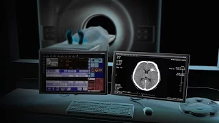
A computed tomography scan (usually abbreviated to CT scan; formerly called computed axial tomography scan or CAT scan) is a medical imaging technique used to obtain detailed internal images of the body. The personnel that perform CT scans are called radiographers or radiology technologists. CT scanners use a rotating X-ray tube and a row of detectors placed in a gantry to measure X-ray attenuations by different tissues inside the body. The multiple X-ray measurements taken from different angles are then processed on a computer using tomographic reconstruction algorithms to produce tomographic (cross-sectional) images (virtual "slices") of a body. CT scan can be used in patients with metallic implants or pacemakers, for whom magnetic resonance imaging (MRI) is contraindicated. Since its development in the 1970s, CT scanning has proven to be a versatile imaging technique. While CT is most prominently used in medical diagnosis, it can also be used to form images of non-living objects. The 1979 Nobel Prize in Physiology or Medicine was awarded jointly to South African-American physicist Allan MacLeod Cormack and British electrical engineer Godfrey Hounsfield "for the development of computer-assisted tomography". (Wikipedia).
CAT or CT scans are used to achieve high resolution images inside the body. But how do they work? Watch the video to find out See more Medical imaging techniques here - https://www.youtube.com/playlist?list=PL5Tk152XlY6SHAnblHcXldLZvYtKUCAUw See www.physicshigh.com for all my videos and
From playlist Medical Physics
CT (Computed Tomography) Scans - A Level Physics
A basic description of the mechanism of CT (computed tomography) scans for medical use in remote sensing. Part of the A Level Physics revision series.
From playlist A Level Physics Revision
Visit our website to learn more using Nucleus content for patient engagement and content marketing: http://www.nucleushealth.com/ This 3D animation created by Nucleus Medical Media presents an overview of how a computerized tomography (CT) machine works and explains why CT scans are taken
From playlist Nucleus Medical Animations in English
ultrasound - A scans explained
Reviews how an A amplitude (A) scan is produced in the context of ultrasound/sonograms See www.physicshigh.com for all my videos and other resources. If you like this video, please press the LIKE and SHARE with your peers. And please add a COMMENT to let me know I have helped you. Follow
From playlist Medical Physics
http://www.nucleushealth.com/ - This 3D medical animation shows the anatomy and physiology of skin and demonstrates an excisional biopsy a surgical techniques commonly used to obtain a sample tissue of suspected skin disorder. A skin biopsy is used to view under a microscope and make a dia
From playlist Healthcare Patient Education Animations
ultrasound/sonogram - B scans explained
Reviews how an a brightness (B) scan is produced in the context of ultrasound/sonograms See www.physicshigh.com for all my videos and other resources. If you like this video, please press the LIKE and SHARE with your peers. And please add a COMMENT to let me know I have helped you. Follow
From playlist Medical Physics
How do PET scans work to detect things such as cancer?
In this video, I discuss Positron Emission Tomography. In particular, I refer to the source of the positron, its annihilation and how the resultant gamma rays are collected. I also briefly discuss its diagnostic benefits. See more Medical imaging techniques here - https://www.youtube.co
From playlist Medical Physics
In this video I show you how to conduct a t-test, analysis of variance, and linear regression in SPSS.
From playlist Healthcare statistics with SPSS
Introduction To Vulnerability Scanning
In this video, I will be introducing you to the process of performing vulnerability scanning, the role it plays in a vulnerability management program, and how it can be used in a penetration test. Vulnerability Scanning is the process of scanning and identifying vulnerabilities, misconfigu
From playlist Nessus
Diagnosing strokes with imaging CT, MRI, and Angiography | NCLEX-RN | Khan Academy
Visit us (http://www.khanacademy.org/science/healthcare-and-medicine) for health and medicine content or (http://www.khanacademy.org/test-prep/mcat) for MCAT related content. These videos do not provide medical advice and are for informational purposes only. The videos are not intended to
From playlist Circulatory system diseases | NCLEX-RN | Khan Academy
3D Image Classification from CT Scans - Keras Code Examples
This video shows you how to use 3D Convolutions to process Viral Pneumonia detection from CT Scans! 3D Image Classification: https://keras.io/examples/vision/3D_image_classification/ Keras Examples: https://keras.io/examples Thanks for watching! Please subscribe and check out the Keras C
From playlist Keras Code Examples
How Technology Gives Insight into Human Anatomy and Disease
(January 12, 2010) Geoffrey D. Rubin, M.D., Stanford Professor of Radiology and Diagnostic Radiology, discusses the various radiology technologies, their uses, and the positive changes they are making in medical diagnosis. Stanford Mini Med School is a series arranged and directed by S
From playlist Lecture Collection | Mini Med School
Ivana Išgum: "Deep learning for cardiovascular image analysis"
Deep Learning and Medical Applications 2020 "Deep learning for cardiovascular image analysis" Ivana Išgum - Amsterdam University Medical Center Abstract: Cardiovascular disease (CVD) is the leading cause of morbidity and mortality worldwide. Medical imaging plays a crucial role in the de
From playlist Deep Learning and Medical Applications 2020
A Blood Test for Brain Damage, and AI Eye Doctors
This week the FDA approves the first ever blood test for diagnosing concussions, and a group of scientists develop a neural network that could save you a trip to the eye doctor. Hosted by: Hank Green ---------- Support SciShow by becoming a patron on Patreon: https://www.patreon.com/scish
From playlist Uploads
Can deep learning AI help with detecting COVID-19/coronavirus?
Join us live where we will have a live paper discussion session on the recent paper on using deep learning to detect COVID-19 from CT lung scan. For slides, paper and more details, visit https://aisc.ai.science/events/2020-03-04/ Papers to be discussed: The Lancet: Feasibility of contro
From playlist Machine Learning for Scientific Discovery
Adam Savage's Curta Calculator Gets CT Scanned!
What's the inside of a Curta mechanical calculator look like? Or how about a Perfect Grade Gundam model? We meet the team at Lumafield, who've created a CT scanner and software that can capture and generate detailed interior models of handheld objects without any physical disassembly. Adam
From playlist Toys, Models and Collectibles
Grave Secrets of Dinosaurs | National Geographic
Paleontologist Phillip Manning shares the exciting cold case of a 67-million-year-old dinosaur — one of the rarest specimens ever unearthed. ➡ Subscribe: http://bit.ly/NatGeoSubscribe About National Geographic: National Geographic is the world's premium destination for science, exploratio
From playlist National Geographic Live!: Season 1
Live from the ATLAS Experiment
Find out more about the ATLAS Experiment and recent upgrades of the detector.
From playlist 360° videos
Randomized Controlled Trials and Full Body CT Scans
Never disrespect the randomized controlled trial. This is Healthcare Triage News. John Green -- Executive Producer Stan Muller -- Director, Producer Aaron Carroll -- Writer Mark Olsen -- Graphics http://www.twitter.com/aaronecarroll http://www.twitter.com/crashcoursestan http://www.twit
From playlist Healthcare Triage News